Tiny Window Transforms Standard Ultrasound Probe into Hi-Res, 3D Imaging Tool
Michaela Martinez
2/11/25Pratt School of Engineering
One small slit could make rapid and detailed imaging tools more accessible for labs and hospitals
SHARE
Tiny Window Transforms Standard Ultrasound Probe into Hi-Res, 3D Imaging Tool
Biomedical engineers have developed a new approach that transforms off-the-shelf, 2D photoacoustic imaging tools into devices capable of rapidly creating accurate and high-resolution 3D images.
In a proof of concept, the team showed that their new approach could show how “forever chemicals” like PFAS affect the early development of embryos in mice.
The research appeared January 29 in the journal Nature Communications.
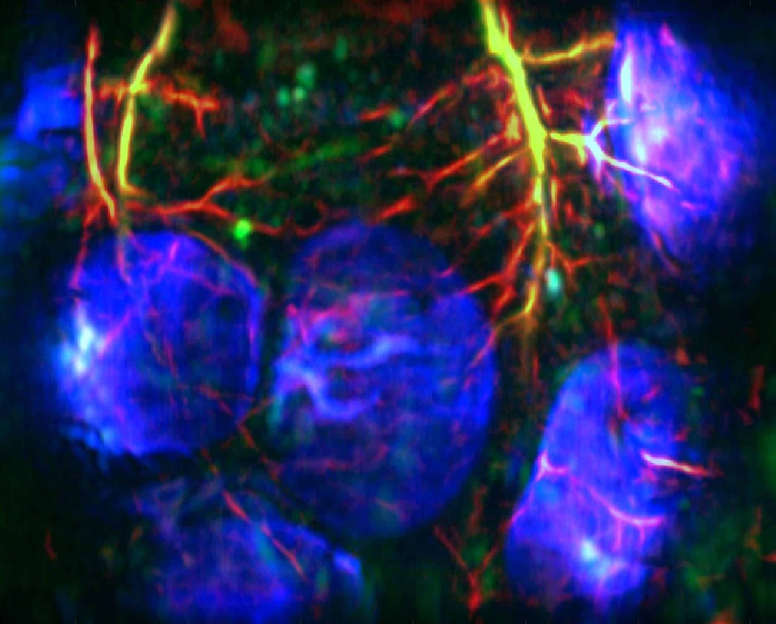
Like the name implies, photoacoustic imaging uses both light and sound to create images of living tissues. The non-invasive imaging technique uses short laser pulses to ‘excite’ the target tissue, releasing ultrasonic waves that are used to create colorful molecular and structural images of deep tissue.
Although photoacoustic imaging is both fast and accurate, access to the tools required to use the technique have stymied its broader use. One of the primary problems involves the ultrasound sensors themselves. Some of the most sophisticated sensors can create detailed 2D and 3D images, but they are expensive and difficult to use.
As a result, most researchers use linear-array transducers, which are the traditional handheld ultrasound sensors used in hospitals. Although these tools are both affordable and widely available, they can only send out and receive ultrasound waves in a single plane. As a result, they lack sensitivity, and the 2D images they produce are poor.
The dilemma is that if you have a good system, it’s expensive, but if you want to cut the price, the image performance dramatically decreases. It’s hard to find the balance, and sometimes that gives people the wrong impression about the viability of this technology.
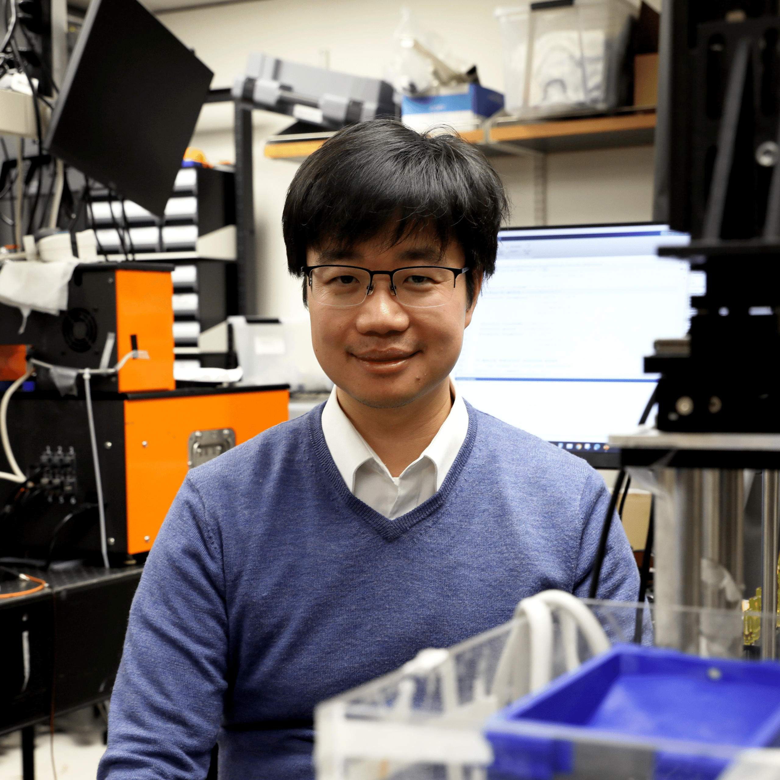
Junjie YaoAssociate Professor of Biomedical Engineering
To make photoacoustic tomography more accessible, Yao and his team devised a deceptively simple solution to improve the sensor. Dubbed 3D diffractive acoustic tomography (3D-DAT), the new system involves placing a cover over their photoacoustic sensor with a small slit for light and soundwaves to enter and exit. When sound waves pass through the opening, they bend and spread out, just as ocean waves move around an obstacle or someone’s voice bends around a corner. The same phenomenon occurs as the ultrasound waves pass back through the slit and hit the sensors. The concept, called diffraction, enables the waves to hit a larger area but doesn’t significantly weaken their energy.
“It’s a bit counter-intuitive, because if you look through a tiny slit, your view decreases,” said Yao. “But diffraction through the slit allows the sound waves to spread a lot more, and the bigger you can spread the waves, the clearer you can see.”
The small change has big results. 3D-DAT enables researchers to transform the 2D, low-resolution transducers into tools that can capture precise, high-resolution 3D data. They paired this advancement with a new graphics processing unit, which uses 3D-DAT’s data to create detailed and colorful 3D images over fifty times faster than previous systems.
“Our small gadget can convert a low-price, bad performance system into a low-price, high-performance system,” said Yao.
The team explored how 3D-DAT performed on experiments they weren’t previously able to conduct using the standard linear-array transducer. These tests included mapping how the molecules that bind to biliverdin––a green pigment––are distributed in glassfrogs and monitoring the circulation and accumulation of gold nanostar particles in a mouse tumor model.
But the most significant results came from their final test, which included studying how per- and polyfluoroalkyl substances, or PFAS, affect embryo development in mice models. Also known as ‘forever chemicals,’ PFAS are found in numerous industrial and consumer products, and they are known to accumulate in living organisms and cause adverse health effects. Using a mouse model that had similar levels of PFAS exposure as a human, they saw that oxygen levels increased during early embryo development, slowing down the growth of blood vessels and negatively affecting embryonic growth and development, especially brain development.
“Our lab found that perinatal exposure to a PFAS mixture comparable to levels found in drinking water in Pittsboro, NC, can adversely affect neurobehavioral development in offspring,” said Dr. Liping Feng, an associate professor of obstetrics and gynecology at the Duke University School of Medicine. “By linking blood oxygen levels and vessel development to PFAS exposure, we offer a concrete, measurable pathway to understanding how these chemicals disrupt normal development.”
“Photoacoustic imaging is getting to a point where we need to figure out how we can broaden the uses and accessibility of this technology so we can make it more mainstream,” said Yao. “We’re hopeful that this new approach will bring the technology into clinics and research labs where scientists can use it to explore these kinds if important medical questions.”
This website uses cookies as well as similar tools and technologies to understand visitors' experiences. By continuing to use this website, you consent to Duke University's usage of cookies and similar technologies, in accordance with the Duke Privacy Statement.I Accept